


Fundus Photography
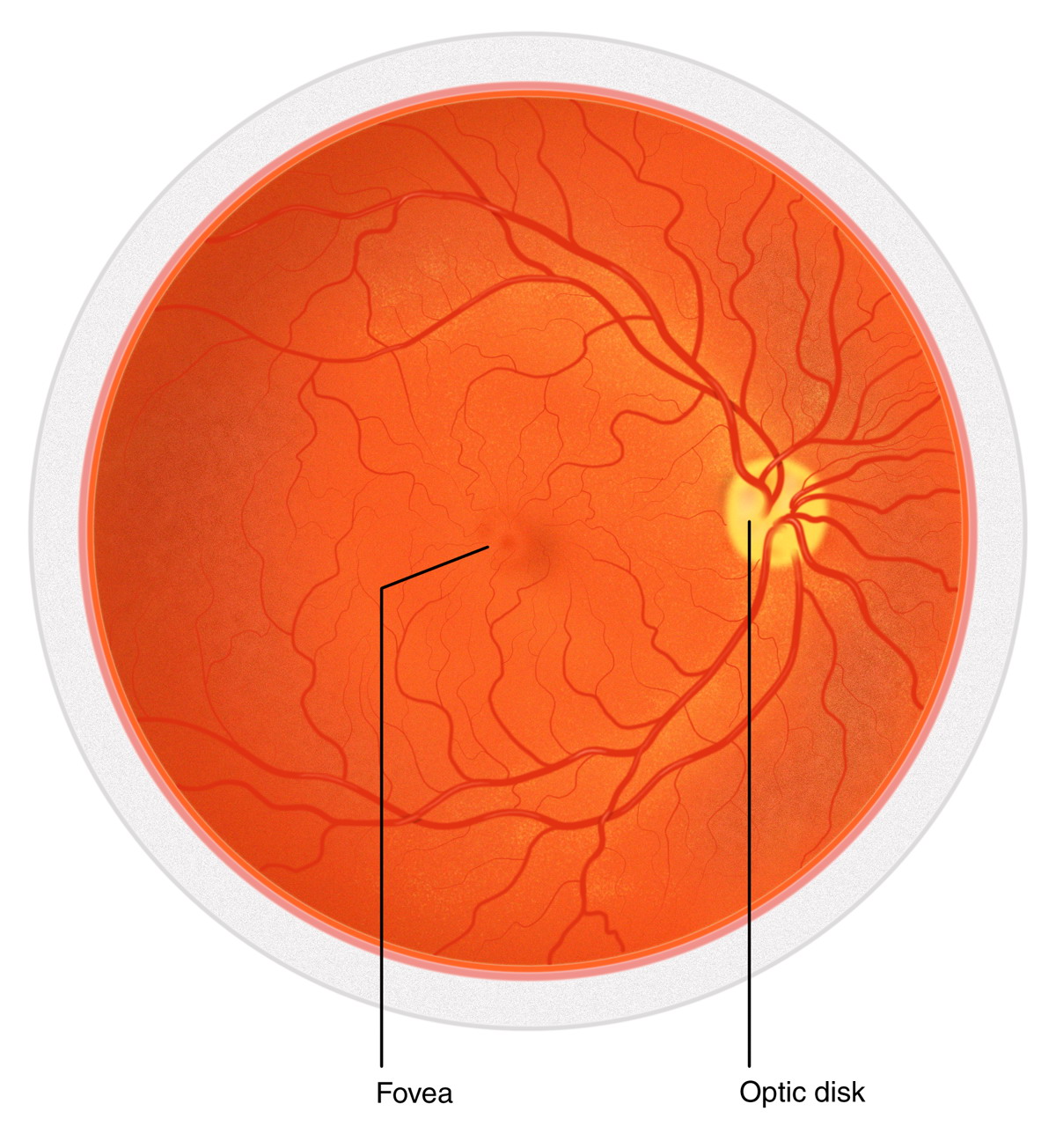 A fundus photograph is a specialized form of medical imaging. Using a customized camera with high-powered lenses that are mounted to a microscope, photographs are taken of the back of the eye by focusing light through the cornea, pupil and lens. Fundus photographs are used to identify or monitor a wide variety of ophthalmic conditions.
To begin the process, the pupil is dilated with eye drops. The patient will be asked to stare at a fixed device, keeping the eyes focused and still. There will be a series of flashes of light. The process usually takes no more than 10 minutes.
Some of the ophthalmic conditions fundus photography is used for include:
- Glaucoma
- Diabetic retinopathy
- Macular edema
- Microaneurysm
- Optic nerve
Fundus photography has also been used to interpret the results of a fluorescein angiogram.
Fluorescein & Indocyanine Green Angiography
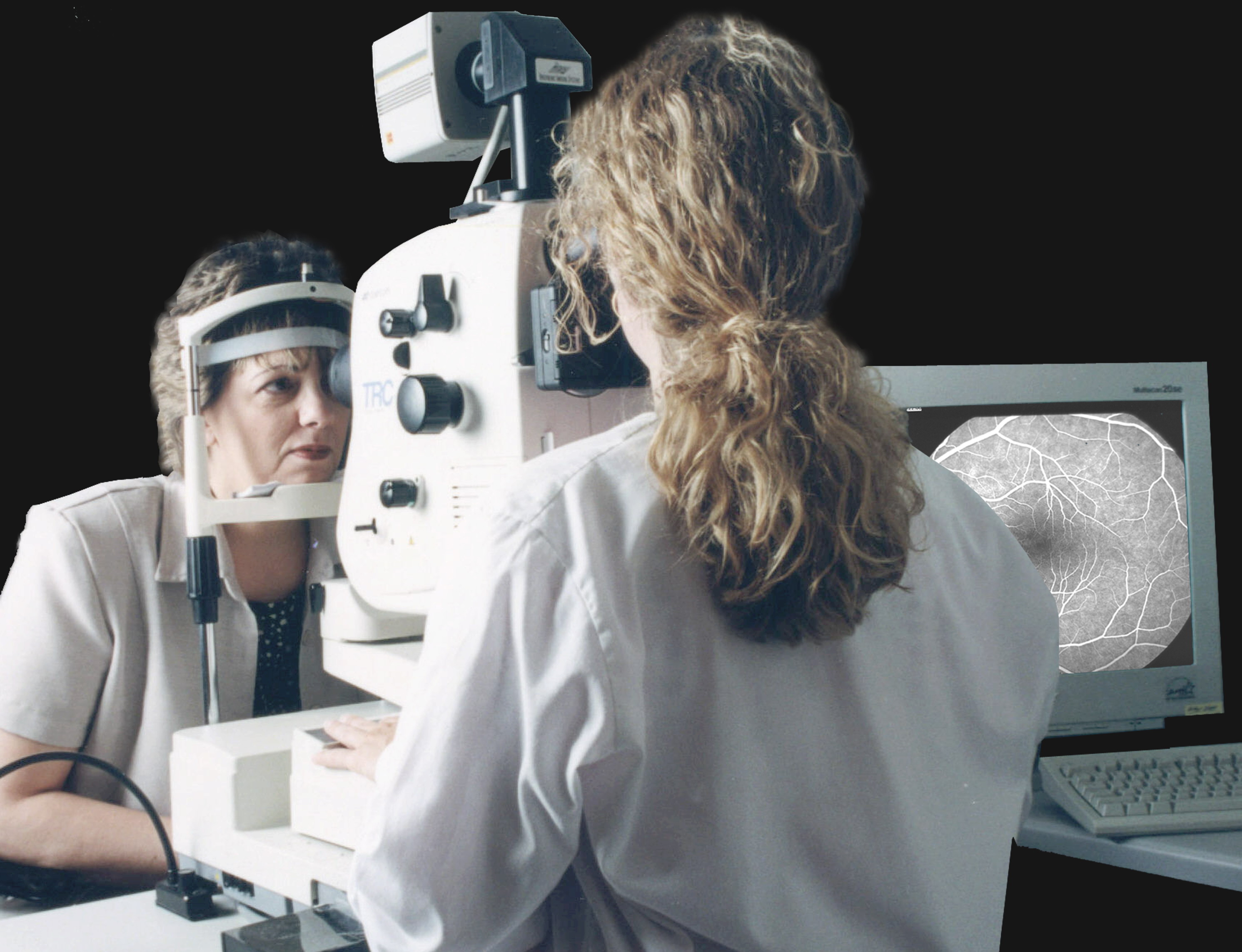 Fluorescein and indocyanine green angiography are diagnostic tests that involve taking photographs of the blood vessels in the eye with the help of a contrast dye. Fluorescein is a yellow dye that allows the eye to glow in visible light, while indocyanine is a green dye that works with infrared light to be captured by the special camera. The images produced by these tests help doctors evaluate the retina and diagnose or monitor problems such as diabetic retinopathy, macular degeneration, abnormal vessel growth, swelling, leaking, retinal detachment, cancer or tumors.
During these exams, the patient's pupils are dilated with eye drops. A few photographs are then taken with a special ophthalmic camera. Next, the contrast dye is injected, usually into the patient's arm. The dye travels up to the eye within a few seconds and "lights up" the blood vessels for the camera. Once the dye is in place, the doctor will take more photographs. The needle is then removed and a final set of photographs is taken approximately 20 minutes later for comparison.
These tests are considered safe for most patients, although it is possible to have a reaction to the dye and develop itching, nausea and a rash. These symptoms can usually be managed through oral medication.
Ocular Ultrasound
Ocular Ultrasound is a high-frequency ultrasound that produces a cross-sectional image of the eye and the space behind the eye.
The Ocular Ultrasound enables eye care professionals to accurately diagnose and evaluate a wide range of eye conditions including:
- Cataracts
- Retinal detachment
- Tissue damage
- Inflammation
- Vitreous Hemorrhage
- Tumor
- Suspected foreign body
- Eye trauma
- Eye pain
- Lens dislocation
Advantages of the Ocular Ultrasound
The Ocular Ultrasound provides numerous advantages over comparable ocular imaging systems. They include:
- Images of the eye can be obtained from all patients
- Accurate results
- Painless procedure
The accuracy of Ocular Ultrasound imaging allows eye care professionals to precisely determine the severity of the patient's condition, ensuring that patients receive the care they need.
Optical Coherence Tomography
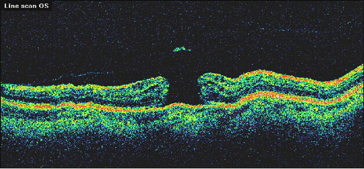
Conditions Detected With an OCT
The images can help with the detection and treatment of serious eye conditions such as:
- Macular hole
- Macular swelling
- Optic nerve damage
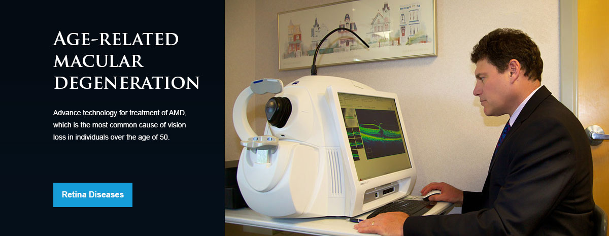
- Age-related macular degeneration
- Macular pucker
- Glaucoma
- Cataracts
- Diabetic eye disease
- Vitreous hemorrhage
OCT uses technology that is similar to that of a CT scan of internal organs. With the scattering of light it can rapidly scan the eye to create an accurate cross-section. Each layer of the retina can be evaluated and measured and compared to normal, healthy images of the retina.
The OCT exam takes about 10 to 20 minutes to perform in your doctor's office, and usually requires dilation of the pupils for the best results.
Photodynamic Therapy
Photodynamic therapy is used to treat a complication of wet macular degeneration in which leaks form beneath the retina. These leaks occur in a structure in the eye called the choroidal neovascular membrane, or CNVM. During photodynamic therapy, dye is injected into the CNVM and a narrow-wavelength laser beam is focused on it for about 90 seconds. The dye absorbs the energy and slows or stops leakage by creating blood clots and stopping abnormal blood vessels from growing. Following treatment, patients should avoid direct exposure to sunlight for several days. Patients most likely to benefit from treatment will have newly onset macular degeneration and no scarring. Vision stabilization is maximized with a series of treatments over one to two years.