

Intravitreal Injections
Intravitreal injections are commonly used to treat retinal diseases such as diabetic retinopathy, macular degeneration and retinal vein occlusion. These diseases often cause blindness and should be treated as early and as thoroughly as possible. FDA-approved medications such as Lucentis, Avastin or Macugen are injected directly into the eye to help patients maintain their baseline vision and keep vision loss at a minimum. Many patients often see an improvement in their vision from these injections as well.
Intravitreal injections are especially effective in treating wet age-related macular degeneration, which, although less common than the dry form, accounts for more than 90% of blindness caused by the disease.
This procedure is performed in a doctor's office and requires only a local anesthetic. Before the medication is injected, the eye is numbed with anesthetic eye drops to help minimize discomfort. The eye is then cleaned with an antiseptic solution and held open with a wire speculum. The medication is then injected directly into the eye. Intravitreal injections are usually administered once a month to maintain eye health in patients with degenerative eye diseases.
Patients may experience some pain or scratchy sensations after the injection. Although rare, side effects may include eye pain, conjunctival hemorrhage, floaters, increased eye pressure and inflammation of the eye.
Retinal Laser Photocoagulation
Retinal laser photocoagulation is a minimally invasive procedure used to seal or destroy leaking blood vessels in the retina that lead to serious retinal conditions such as diabetic retinopathy and macular edema. This procedure can also seal retinal tears and destroy abnormal tissue found in the back of the eye.
Pneumatic Retinopexy
Pneumatic retinopexy is a surgical procedure used to repair some forms of retinal detachment. It is an effective treatment method when the detachment is in the upper portion of the retina and has been caused by one tear or several small tears in one location.
A pneumatic retinopexy is typically performed on an outpatient basis using local anesthesia. A gas bubble is injected into the eye in order to provide mild pressure on the retina and flatten it. This allows any fluid trapped beneath the detachment to be withdrawn. Either laser photocoagulation or cryotherapy will be used to close the retinal tear.
The gas bubble remains in the eye for a few weeks until it is naturally absorbed. It continues to gently compress the retina, which helps to seal the retina in position. During this recovery period, it is important to follow your surgeon's instructions on head and eye positioning. This keeps the gas bubble in the right place and prevents it from traveling within the eye. You will be restricted from lying on your back and riding in an airplane for several weeks to avoid complications arising from gas bubble movement.
Pneumatic retinopexy has a high success rate for retinal reattachment. Vision can often be restored, particularly if the macula was not involved.
Retina Reattachment Surgery
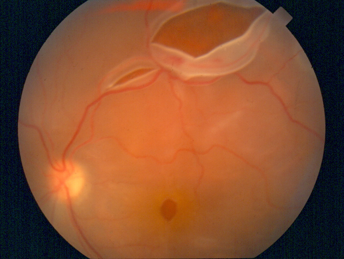 Retinal detachment occurs when the retina of the eye is pulled away from the underlying tissue to which it is attached. A retinal detachment is a medical emergency which can lead to permanent blindness if left untreated. In most cases, the detachment is a slowly progressing issue which must be treated once symptoms are realized. In some cases, a detachment occurs due to a trauma which causes a tear in the retina, allowing fluid to enter the vitreous and pull on the retinal tissue.
Symptoms of a Retinal Detachment
Symptoms of retinal detachment may progress slowly or rapidly, but should be reported to a doctor as soon as possible so as to minimize the risk of vision loss. Some of the symptoms of a retinal detachment include:
- A dense shadow throughout the visual field
- A sudden decrease in visual acuity
- A sudden increase in the amount of "floaters" in vision
- Bright flashes in the periphery
- An unnatural "curving" of straight lines
- Loss of central vision
Treatment for a Retinal Detachment
There are several treatment options available for retinal detachment, depending on the severity of the condition and overall health of the patient. Patients with small tears can usually be treated through laser surgery or a freeze treatment called cryopexy. Some patients may require a small incision retinal surgery known as victrectomy to remove the vitreous and move the retina back in place.
Most surgeries to repair a retinal detachment are successful. In some cases, a second procedure will need to be performed. After a successful procedure, vision will take time to improve but may not return to previous levels of acuity.